

Since the circumcised area of a tumor is less rigid than the tumor itself, it is possible to detect them in soft tissues like breasts or skin, using methods such as ultrasound or magnetic resonance imaging. Furthermore, this fact has led to the development of devices with tunable viscoelastic properties capable of changing cell behavior, as well as mechanotransduction systems. These devices work by initiating intracellular signaling pathways and causing the activation of ion channels or protein kinases, and even changing gene transcription, protein production, phenotype and altering tumor progression.
Researchers at University of Granada (UGR) have developed a non-invasive medical device that generates shear at a controlled frequency and amplitude to impact the mechanotransduction pathway in a way that influences cell proliferation and, consequently, tumor growth.
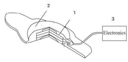
The invention describes a device that emits shear waves and deposits a minimal amount of energy on the tissue. This procedure is performed through the surface of a patient to a specific region of cancer cells, which is located at a specific depth beneath the skin. The aim is to aid classical tumor therapy for instance by rendering cells more receptive to drugs, or by gaining time during chemotherapy by reducing cell migration and, hence slowing down the metastatic process. The device consists of a piezoelectric part (1) consisting of electrodes, attenuating backing and acoustic lens in contact with the patient’s body, a cage (2) housing the receiver, and external electronics (3).
The benefits have been thoroughly tested in different studies. One of them targeted cancer stem cells (CSC) using axisymmetric shear waves in vitro for effectiveness with different frequencies and amplitudes of torsional waves.
Benefits:
- The vibrating shall have a minimal surface area that enables the causing of mechanotransduction effects for cells within the target zone while leaving cells outside of the target zone unaffected.
- The transducers are operable for functioning within and outside of the main magnetic field. This enables the image to be removed from the magnetic resonance imaging system.
- It measures the anisotropy of soft biological tissue to characterize tissue parameters and know how to influence cell proliferation and tumor growth.
- It is a disruptive technology that allows the reduction of doses and the duration of the chemotherapeutic treatment, with a subsequent reduction of mortality and adverse effects.
- Reduction of health costs.
The represented institution is looking for a collaboration that leads to commercial exploitation of the presented invention.
Institution: Universidad de Granada
TRL: 3-4
Contact: Noelia Mas / noelia@viromii.com